Microscopic spinal fixation is a minimally invasive surgical procedure performed using the latest technology to surgically stabilize spinal segments with plates, screws, cages, and more. Dr. Zeiad Yossry, Professor of Neurosurgery and Parkinson's Disease, performs this minimally invasive procedure through a smaller incision than traditional surgery to achieve the same result. The surgery utilizes a highly sensitive surgical microscope, specialized surgical instruments, and advanced imaging techniques to visualize and perform the procedure through small incisions.
Vertebral column:
The spine plays a vital role in providing stability, smooth movement, and protection for the delicate spinal cord. The spine is made up of bony structures called vertebrae, stacked on top of each other, separated by fibrous tissues called intervertebral discs. The vertebrae and discs form the spinal column from the neck to the pelvis, providing coordination and support to the body.
Age-related changes, trauma, or medical conditions may cause your vertebrae to shift from their normal position, affecting the surrounding nerves, muscles, and ligaments, leading to severe pain and discomfort.
Indications for spinal stabilization:
Dr. Zeiad Yossry recommends spinal stabilization surgery when there is trauma or instability in a part of the spine, or when spinal surgeries may lead to instability over time. Microsurgical spinal stabilization is a useful procedure for treating various types of spinal disorders, including:
• Spinal fractures.
• Spinal cord tumors.
• Spinal deformity.
• Degenerative disc disease.
• Spinal canal stenosis.
• Herniated discs.
What happens during surgery?
Spinal fusion is used to fuse vertebrae together or to stabilize a vertebra and its adjacent disc. The procedure aims to create a solid structure between adjacent vertebrae, eliminating movement between them.
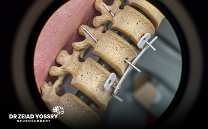
Damaged vertebrae are fused using spacers, bone grafts, metal rods, and screws. Screws are anchored to the spine and connected to rods to fuse the spine. Bone grafts may also be placed along the corrected spine. These grafts do not fuse at the time of surgery but provide the foundation and environment for new bone to grow and integrate the vertebrae.
Goals of spinal fusion surgery:
1. Improve spinal alignment by correcting any deformity (e.g., forward or lateral slippage of vertebrae).
2. Release any trapped nerves.
3. Remove the source of the pain - the degenerated and inflamed disc.
4. Disc fusion by filling the space between the vertebrae with a cage containing a graft to stimulate bone growth and ultimately fuse the vertebrae.
Benefits of microscopic spinal fusion:
The microscope magnifies the surgical site, allowing clear visualization of the disc and surrounding nerves. The procedure uses an integrated 3D imaging system with intraoperative navigation to precisely place screws and plates, enhancing safety, accuracy, and minimizing muscle and tissue damage. Therefore, microscopic surgery offers several advantages over traditional spinal fixation, including:
• Smaller surgical scars.
• More effective surgical results.
• Reduced risk of infection.
• Less blood loss during surgery.
• Less postsurgical pain.
• Faster recovery.
• Shorter hospital stay.
• Better cosmetic results.
• Quick return to work and normal activities.
Preparing for microscopic vertebral fixation:
• Dr. Zeiad Yossry, Professor of Neurosurgery and Parkinson's Disease, performs a comprehensive examination to check for any medical problems that need to be addressed before surgery.
• Depending on your medical history and age, you may need to undergo tests such as blood tests and imaging to help detect any abnormalities that could threaten the safety of the procedure.
• You will be asked if you are allergic to medications or anesthesia.
• You should refrain from taking medications or supplements such as blood thinners, aspirin, or anti-inflammatory drugs for one to two weeks before surgery.
• You should refrain from smoking for at least a few days before surgery.
• You should not eat any solid foods or liquids for at least 8 hours before surgery.
Microscopic vertebral fixation procedure:
Minimally invasive microsurgical spinal fixation typically involves the following steps:
• You will lie face down on the operating table under general anesthesia.
• Under X-ray guidance, Dr. Zeiad Yossry passes a thin needle called a guide wire through the skin and back muscles to identify the affected area.
• The needle is withdrawn, and a small incision of about 2 cm is made near the targeted vertebra. Expanders are inserted to gently move muscles and other soft tissues aside, creating a tunnel for surgery.
• Once the disc or vertebral bones are exposed, the degenerated disc is removed, and a bone graft or synthetic material is placed into the disc space.
• Through microscopic visualization, the surgeon performs the necessary stabilization. Special rods, screws, plates, cages, or hooks are placed to securely stabilize the spine.
• An intraoperative 3D imaging system with surgical navigation verifies the precise placement of screws and plates, ensuring high safety and accuracy with minimal damage to surrounding tissues, muscles, and nerves.
• After fixation, the microscope and dilators are withdrawn, and the soft tissues are repositioned; then the incision is closed.
• A small adhesive strip is placed over the incision, and the patient is transferred to the recovery area.
Recovery after microscopic vertebral fixation:
In general, the post-operative instructions provided by Dr. Zeiad Yossry, Professor of Neurosurgery and Parkinson's Disease, following microscopic spinal fusion include:
• You will be transferred to the recovery area, where the nurse will closely monitor your vital signs during recovery.
• You may feel mild pain around the incision area. This pain will subside within a few days, and in the meantime, you will be given medication to manage it.
• You will undergo some X-rays or CT scans to assess the outcome of the procedure.
• You may need to stay in the hospital for one or two days before being discharged.
• Walking and gentle moving around in bed is highly recommended to avoid the risk of blood clots.
• Antibiotics are prescribed as needed to combat the risk of infection associated with surgery.
• Your diet will gradually progress after surgery, starting with clear liquids and advancing to solid foods as tolerated.
• Instructions on wound care and bathing will be provided.
• A calcium-rich, low-fat diet is highly recommended to promote faster healing and recovery.
• It is recommended to follow a diet rich in fiber and drink 8 to 10 glasses of water daily to maintain soft bowel movements.
• Activity restrictions until the first follow-up visit include:
o Avoid bending your back.
o Do not lift anything heavier than 2.5 kg.
o Avoid strenuous activities such as housework, yard work, or sexual intercourse.
Risks and complications:
Microscopic vertebral fixation is considered a relatively safe procedure; however, as with any surgical procedure, some risks and complications may occur, such as:
• Infection.
• Bleeding.
• Blood clots.
• Allergic reactions.
• Neurovascular injury.
• Persistent pain.
• Bowel or bladder problems.
Spinal stabilization does not significantly limit spinal movement or daily activities; on the contrary, it ensures the integrity of the spine. Some patients mistakenly believe that stabilization causes limitations during movement or bending, but this is not true. Other vertebrae compensate for the movement lost due to fusion, allowing patients to live normally—especially when performed by a skilled and experienced surgeon, such as Dr. Zeiad Yossry, Professor of Neurosurgery and Parkinson's Disease, who makes precise surgical decisions. Also, determining the appropriate size and orientation of the screws requires his unique expertise to avoid any injury.
Microscopic spinal fixation is an effective surgical solution for treating spinal problems while maintaining minimal surgical invasiveness. Thanks to Dr. Zeiad Yossry's expertise, this surgery is performed with high precision to achieve the best outcomes and minimize complications, offering patients a pain-free life and a quick return to normal activity.