Microdiscectomy is a minimally invasive surgical procedure used to treat herniated discs. When a herniated disc compresses a spinal nerve, symptoms may include pain (which may radiate down the arms and legs, or both, as in sciatica), muscle weakness, and moving difficulties.
In the past, traditional discectomy required a large incision and complete removal of the disc. Today, with advanced techniques and equipment, Dr. Zeiad Yossry, Professor of Neurosurgery and Parkinson's Disease, performs microscopic discectomy, often providing rapid or even immediate pain relief.
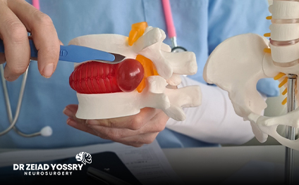
What is a herniated disc?
A healthy intervertebral disc contains a soft, gelatinous center (nucleus) surrounded by a rubbery outer layer (annulus). A herniated disc, also known as a bulging disc or slipped disc, occurs when part of the nucleus protrudes through a tear in the annulus. This often results from age-related wear and tear, known as disc degeneration. As we age, these discs become less elastic and are more prone to tearing or herniation, even with minor stress.
Although a herniated disc can occur in any part of the spine, it is most common in the lower back (lumbar spine) and less common in the neck (cervical spine). If left untreated, a herniated disc can irritate a nearby nerve, causing pain, weakness, or tingling that radiates from the back to the buttocks and down the leg.
What is microdiscectomy?
Microscopic discectomy is a surgical procedure to relieve pain and other symptoms caused by a herniated disc pressing on a nearby nerve root. During the procedure, Dr. Zeiad Yossry, Professor of Neurosurgery and Parkinson's Disease, relieves the nerve by removing small portions of the disc, bone, and ligaments.
Microdiscectomy is considered a minimally invasive procedure because it requires only a small incision and a microscope to magnify the surgical site. The surgeon also uses smaller instruments and devices to operate within the limited space of the spine.
How is microscopic discectomy performed?
Small incisions are made over the affected area of the spine. An illuminated surgical microscope helps Dr. Zeiad Yossry visualize the herniated disc and surrounding tissues, allowing precise removal of the herniated disc portion beneath the nerve root. By creating more space, pressure is relieved, enabling the nerve root to heal.
Because microscopic discectomy involves small incisions, patients often experience less pain, fewer complications, and reduced trauma to surrounding tissues, leading to faster recovery and an earlier return to daily activities. Additionally, the risk of disc re-herniation is lower.
When does Dr. Zeiad Yossry recommend microscopic discectomy?
Dr. Zeiad Yossry, Professor of Neurosurgery and Parkinson's Disease, recommends microdiscectomy after conservative treatments, including physical therapy, corticosteroid injections, and medications, have been tried for at least 6–12 weeks without any relief.
In some cases, poor mobility may be a reason for earlier surgical intervention. Patients with cauda equina syndrome require immediate surgery, as nerve compression in the lower spine affects bladder and bowel function.
What are the benefits of microscopic discectomy?
The surgical goal is to remove the disc fragment and any bone or ligament that compresses the nerve root. To achieve this, Dr. Zeiad Yossry creates a small window in the vertebra, removes the herniated portion, and relieves nerve compression.
Imaging techniques, including X-rays, are used before and sometimes during surgery to ensure the correct site is treated.
Advantages of microscopic discectomy:
The microscope is a multidimensional imaging system used during surgery. It allows Dr. Zeiad Yossry to perform surgery with greater precision in positioning spinal instruments to improve surgical outcomes. Combined with the navigation system, it shortens surgical time, minimizes reoperation risk, and exposes patients to less radiation during imaging. Furthermore, intraoperative neurophysiological monitoring is used to monitor potential nerve damage during surgery, resulting in:
• Reduced damage to surrounding nerves and muscles.
• Smaller incision with better cosmetic result.
• Less pain.
• Faster recovery and quicker return to daily life.
What are the possible complications of microscopic discectomy?
During surgery, multiple safety measures are taken to control bleeding, prevent infection, and avoid injury to surrounding tissues. Rarely, a tear in the tissue surrounding the spinal nerves may occur, which the surgeon repairs using a collagen patch.
What is the expected recovery time after a microscopic discectomy?
After a two-week rest period to allow soft tissues to heal, many patients feel well enough to return to work.
Is microscopic discectomy painful?
After surgery, most patients improve with minimal pain medication and a muscle relaxant. While some discomfort from the surgical incision may occur, many patients experience rapid relief from herniated disc pain.
Pain relief may take longer in patients who have had prolonged nerve compression and associated symptoms. When leg pain is the primary symptom, it usually subsides over time. Additionally, patients may experience temporary muscle cramps, mild numbness, or tingling after surgery, which typically resolves gradually.
Who is eligible for microdiscectomy?
Most patients with a herniated disc who do not respond to medications and physical therapy over time are eligible for a microscopic discectomy. While this condition commonly affects individuals aged 30–50, it can occur outside this age range.
Herniated discs are rare in children and young adults, who tend to recover without surgery. Microscopic discectomy may also be suitable for adults in their 80s or 90s, though they should be cautioned about the increased risk of medical or surgical complications in this age group.
How successful is microdiscectomy?
Overall, the success rates of microscopic discectomy performed by Dr. Zeiad Yossry, Professor of Neurosurgery and Parkinson's Disease, are excellent, with many of his patients expressing a high degree of satisfaction with the outcome. Presurgical evaluation, along with the patient's commitment to maintaining good spinal health after microdiscectomy, contributes to the success of the surgery.
Some individuals with a herniated disc also have other spinal problems that cause nerve-related pain and disability. In such cases, additional procedures may be required on one or more vertebrae if these conditions contribute to their disability.
Can a microdiscectomy be performed for a disc that has been herniated for the second time?
In cases of recurrent herniated discs, a microdiscectomy can be performed on the same disc, often with good results. However, if the herniated disc occurs in the same disc a third time, a different type of treatment will be recommended.
Microdiscectomy with Dr. Zeiad Yossry, Professor of Neurosurgery and Parkinson's Disease, represents an effective solution for treating herniated discs with minimal surgical intervention, allowing for successful results with less pain and a faster recovery. Thanks to this advanced technique and Dr. Zeiad Yossry's expertise, patients can regain their mobility and quality of life with confidence and safety, achieving the best results and the lowest complication rates.